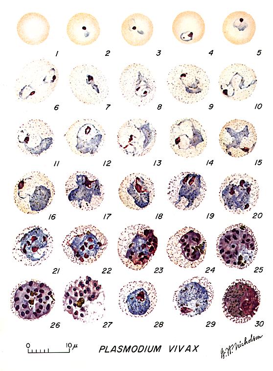
A: Stages of P. vivax in thin smears. Fig. 1: Normal red cell; Figs. 2-6: Young trophozoites (ring stage parasites); Figs. 7-18: Trophozoites; Figs. 19-27: Schizonts; Figs. 28 and 29: Macrogametocytes (female); Fig. 30: Microgametocyte (male).
Illustrations from: Coatney GR, Collins WE, Warren M, Contacos PG. The Primate Malarias. Bethesda: U.S. Department of Health, Education and Welfare; 1971. Reproduced here courtesy of the CDC (www.cdc.gov).
Leave a Reply
You must be logged in to post a comment.